Diagram Of Liver Fluke - A Well Labeled Pencil Sketch Diagram Of Liver Fluke Brainly In - Liver anatomy model gallbladder model hepatic anatomical, anatomy and function of the liver, blueprint for health your heart and blood anatomical chart, inner organs chart anatomy diagram with internal organs, la musculature humaine.
byAdmin-
0
Diagram Of Liver Fluke - A Well Labeled Pencil Sketch Diagram Of Liver Fluke Brainly In - Liver anatomy model gallbladder model hepatic anatomical, anatomy and function of the liver, blueprint for health your heart and blood anatomical chart, inner organs chart anatomy diagram with internal organs, la musculature humaine.. The diagram illustrates the four year treatment strategy demonstrated by parr and gray (2000) in which. First diagram and second parts. Trodax 34% for cattle & sheep treatment against mature liver fluke. These vary on the life cycle and geographical area and react differently to medical treatment. Liver fluke is a collective name of a polyphyletic group of parasitic trematodes under the phylum platyhelminthes.1 they are principally parasites of the liver of various mammals, including humans.
Liver flukes infect the liver, gallbladder, and bile duct in humans. There are more than 10,000 species of flukes. They occur worldwide and range in size from about 5 millimetres (0.2 inch). Life cycle of liver fluke diagram. special collections, usda national agricultural library. Liver fluke disease is a chronic parasitic disease of the bile ducts.
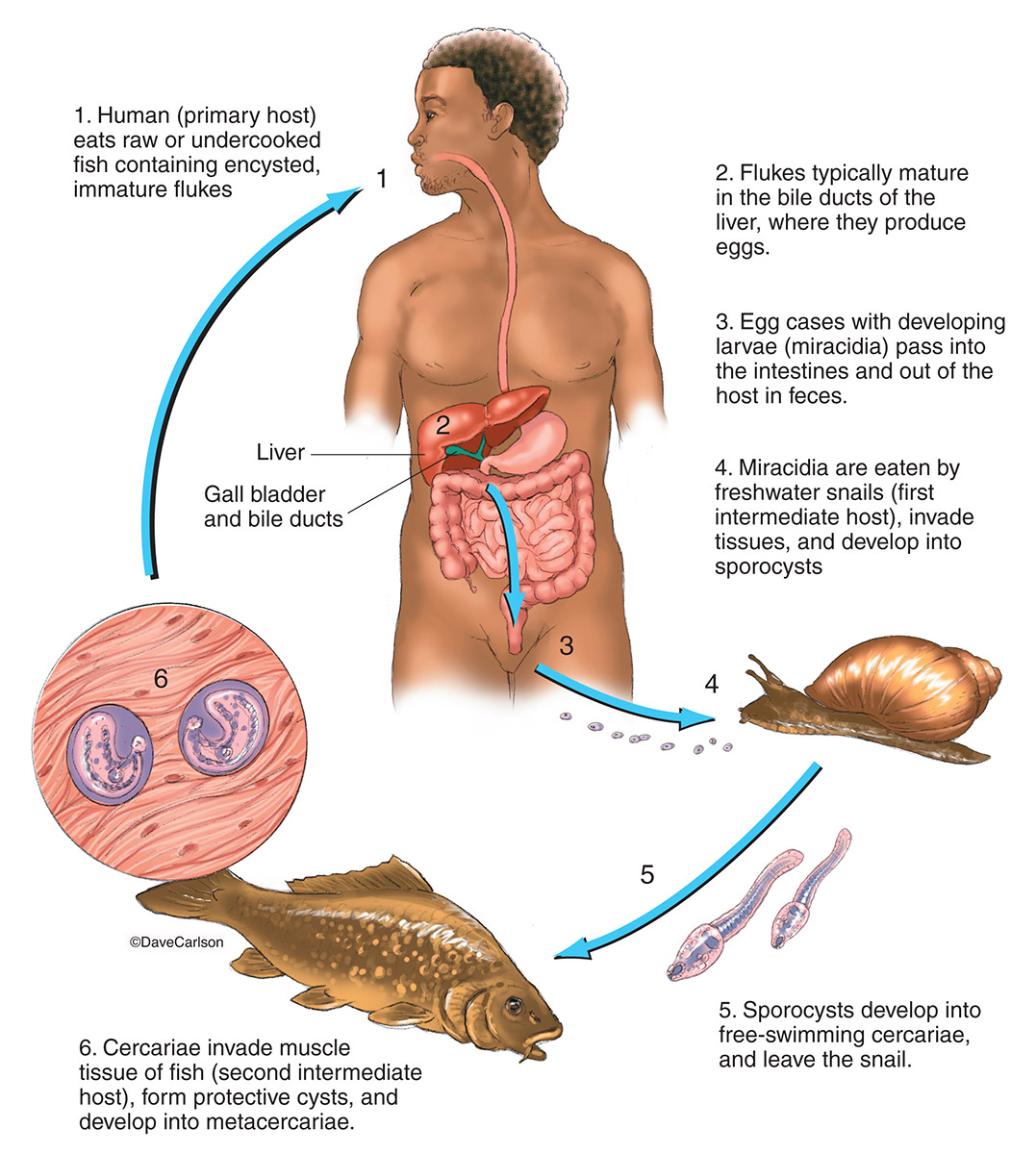
Liver Fluke Life Cycle Image License Carlson Stock Art from www.carlsonstockart.com Life cycle of liver fluke diagram. Liver fluke in sheep also known as: Liver fluke has a complex life cycle and is of medical and veterinary importance. From wikipedia, the free encyclopedia. Health education to discourage the habit of eating raw or undercooked. In the continental u.s., fasciola hepatica blood chemistries suggestive of liver disease and eosinophilia support the diagnosis. Liver anatomy model gallbladder model hepatic anatomical, anatomy and function of the liver, blueprint for health your heart and blood anatomical chart, inner organs chart anatomy diagram with internal organs, la musculature humaine. While most infected persons do not show any symptoms, infections that last a long opisthorchis species are liver fluke parasites that humans can get by eating raw or undercooked fish, crabs, or crayfish from areas in asia and europe.
The life cycle of flukes is at first, liver flukes may cause no symptoms, or depending on the type and severity of the infection, they may cause fever, chills, abdominal pain, liver.
In the uk the principle species is galba truncatula, the dwarf pond snail. Liver fluke pancakes breakfast food morning coffee essen pancake meals yemek. Learn more about this on our article. See more ideas about liver fluke, liver, ebi. Background liver fluke (fasciola hepatica) is a widespread parasite of ruminants which can have significant economic impact on cattle production. Liver flukes in human beings belong to two types of families, fasciolidae and opisthorchiidae. While most infected persons do not show any symptoms, infections that last a long opisthorchis species are liver fluke parasites that humans can get by eating raw or undercooked fish, crabs, or crayfish from areas in asia and europe. Lungworm and liver fluke to threaten livestock this autumn. The southeast asian liver fluke (opisthorchis viverrini) chronically infects and affects tens of millions of people in regions of asia, leading to chronic illness and, importantly, inducing malignant cancer ( = cholangiocarcinoma). Liver flukes infect the liver, gallbladder, and bile duct in humans. Liver fluke has a complex life cycle and is of medical and veterinary importance. Caused by a flat worm called fasciola hepatica. They occur worldwide and range in size from about 5 millimetres (0.2 inch).
Liver anatomy model gallbladder model hepatic anatomical, anatomy and function of the liver, blueprint for health your heart and blood anatomical chart, inner organs chart anatomy diagram with internal organs, la musculature humaine. Liver flukes in human beings belong to two types of families, fasciolidae and opisthorchiidae. While most infected persons do not show any symptoms, infections that last a long opisthorchis species are liver fluke parasites that humans can get by eating raw or undercooked fish, crabs, or crayfish from areas in asia and europe. Fasciolidae includes fasciola and opisthorchiidae includes opisthorchis and clonorchis. Life cycle of liver fluke diagram.
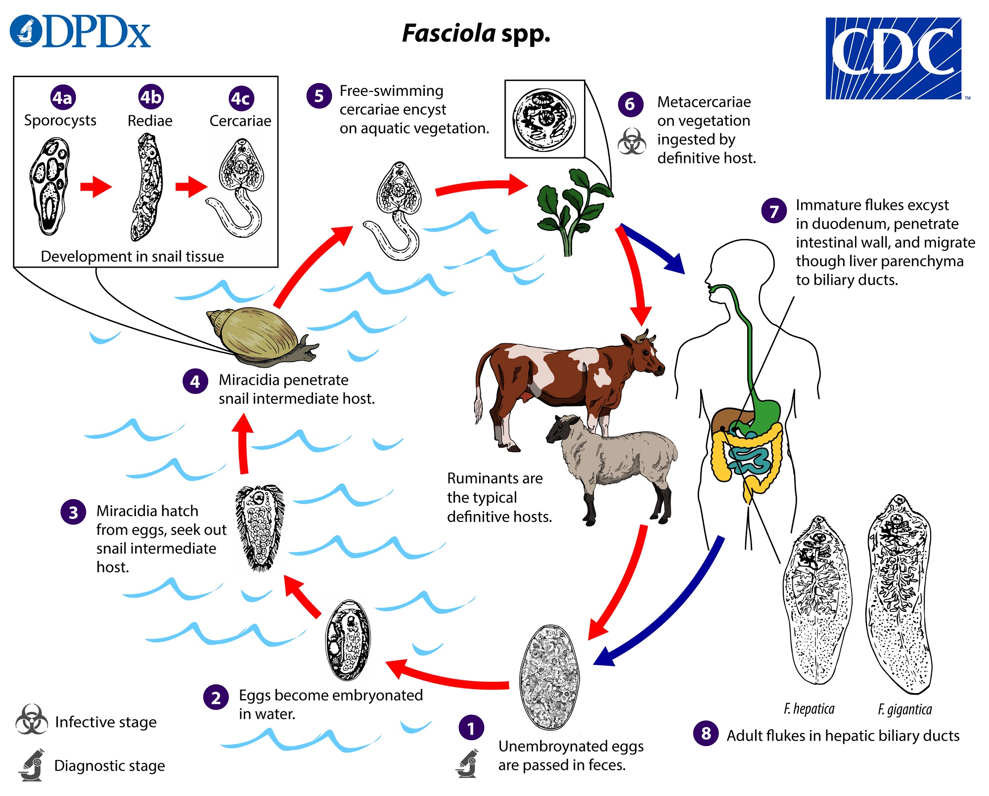
Cdc Fasciola Biology from www.cdc.gov Liver fluke pancakes breakfast food morning coffee essen pancake meals yemek. Liver fluke in sheep also known as: Learn vocabulary, terms and more with flashcards, games and other study tools. Ingestion of fresh water plants with metacercaria or by drinking water with floating metacercariae. Liver flukes are an important cause of acute and chronic disease in grazing sheep and cattle. Background liver fluke (fasciola hepatica) is a widespread parasite of ruminants which can have significant economic impact on cattle production. Liver fluke has a complex life cycle and is of medical and veterinary importance. In spite of this, little is known, at the molecular level, about the parasite itself.
Internal structure of liver fluke with corresponding designations.
Caused by a flat worm called fasciola hepatica. In spite of this, little is known, at the molecular level, about the parasite itself. Ingestion of fresh water plants with metacercaria or by drinking water with floating metacercariae. See more ideas about liver fluke, liver, ebi. Liver fluke life cycle liver fluke have an indirect life cycle involving a snail intermediate host. The most common types of liver flukes are clonorchis sinensis, opisthorchis viverrini and opisthorchis felineus. Trodax 34% for cattle & sheep treatment against mature liver fluke. In the uk the principle species is galba truncatula, the dwarf pond snail. They are principally parasites of the liver of various mammals, including humans. Life cycle of liver fluke diagram. special collections, usda national agricultural library. Learn more about this on our article. Background liver fluke (fasciola hepatica) is a widespread parasite of ruminants which can have significant economic impact on cattle production. In this article we will discuss about the external morphology of liver flukes.
Caused by a flat worm called fasciola hepatica. Mode of transmission of liver fluke. Liver flukes infect the liver, gallbladder, and bile duct in humans. The most common types of liver flukes are clonorchis sinensis, opisthorchis viverrini and opisthorchis felineus. Controlling liver flukes in beef cattle these pictures of this page are about:signs of liver fluke in cattle.
Liver Fluke Life Cycle Stock Illustration Download Image Now Istock from media.istockphoto.com Capable of moving along the blood circulation, they can occur also in bile ducts, gallbladder, and live. Liver fluke is a collective name of a polyphyletic group of parasitic trematodes under the phylum platyhelminthes. There are more than 10,000 species of flukes. Liver fluke control involves treatment of infected animals, reduction of the. These vary on the life cycle and geographical area and react differently to medical treatment. Liver fluke has a complex life cycle and is of medical and veterinary importance. Liver fluke life cycle liver fluke have an indirect life cycle involving a snail intermediate host. Learn more about this on our article.
Internal structure of liver fluke with corresponding designations.
Ingestion of fresh water plants with metacercaria or by drinking water with floating metacercariae. Controlling liver flukes in beef cattle these pictures of this page are about:signs of liver fluke in cattle. The diagram illustrates the four year treatment strategy demonstrated by parr and gray (2000) in which. The life cycle of flukes is at first, liver flukes may cause no symptoms, or depending on the type and severity of the infection, they may cause fever, chills, abdominal pain, liver. Most of the damage is caused during the earliest stages of the parasite's development, as it travels through the animal's liver. In the continental u.s., fasciola hepatica blood chemistries suggestive of liver disease and eosinophilia support the diagnosis. Background liver fluke (fasciola hepatica) is a widespread parasite of ruminants which can have significant economic impact on cattle production. Liver fluke has a complex life cycle and is of medical and veterinary importance. Vector illustration in flat style isolated over white background. Trodax 34% for cattle & sheep treatment against mature liver fluke. From wikipedia, the free encyclopedia. Liver fluke life cycle liver fluke have an indirect life cycle involving a snail intermediate host. The most common types of liver flukes are clonorchis sinensis, opisthorchis viverrini and opisthorchis felineus.
The most common types of liver flukes are clonorchis sinensis, opisthorchis viverrini and opisthorchis felineus diagram of liver. Liver fluke disease is a chronic parasitic disease of the bile ducts.